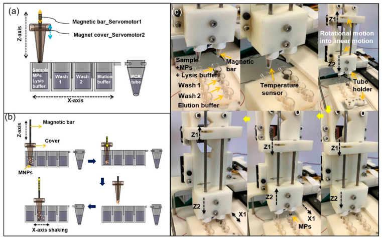
Figure 2.
Schematic images of (a) cross-sectional view of a sample cartridge and (b) automated workflows including mixing, capturing, transport, and dispersion. The cartridge has four wells: lysis buffer, washing I, washing II, and elution buffer for automated sample preparation, and a conical PCR microtube for DNA amplification. (c) Photographs of each step for magnetic particle (MP) transport and re-dispersion to next well.

