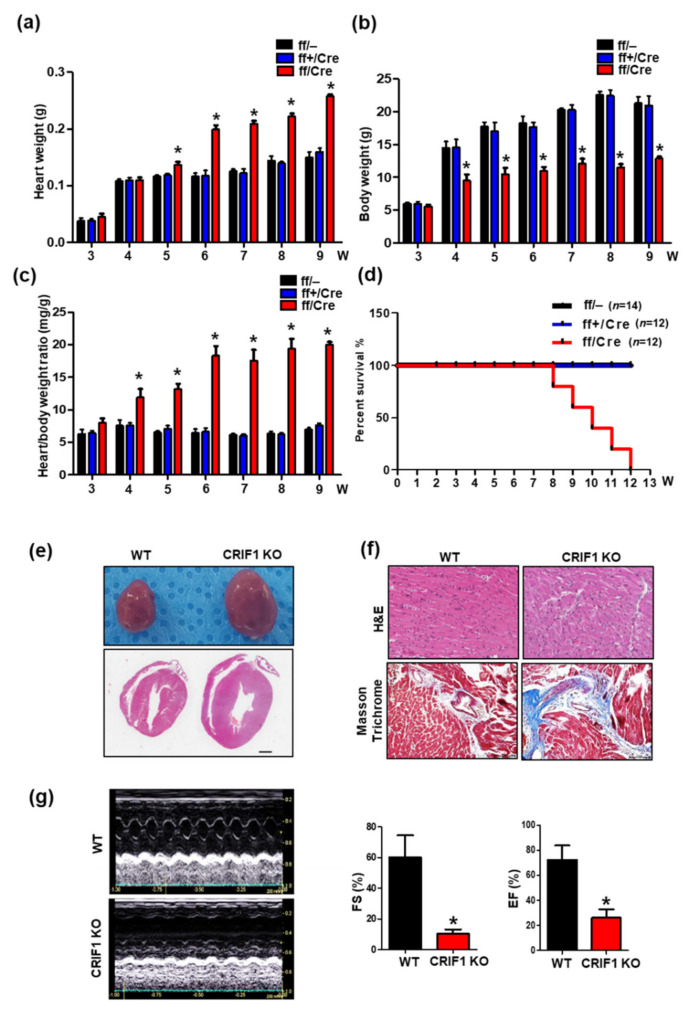
Figure 2.
Endothelial cell-specific deletion of CRIF1 causes cardiac defects. (a) Body weight and (b) heart weight and (c) heart/body weight of 3 to 9-week-old mice (n = 4). (d) Survival rate of WT (ff/–), hetero (ff+/Cre), and CRIF1 EKO mice (ff/Cre) (n = 12~14 mice in each group). (e) Representative whole mount hearts (top) and hematoxylin and eosin staining of four chamber histological sections (bottom) and from the hearts of mice. (Scale bar, 1 mm) (f) Representative H&E staining and Masson’s trichrome staining of high-magnification sections of the hearts of mice. (g) Representative M-mode echocardiograms from WT (up) and CRIF1 EKO (down). Percentage of FS and EF determined from echocardiography (n = 4 mice in each group). All representative images were examined from 9-week-old mice. All data are presented as means ± SEM of at least three independent experiments. * p < 0.05 vs. WT group.

