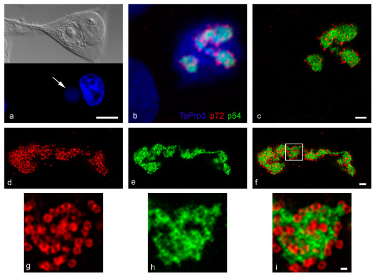
Figure 1.
ASFV infected Vero cells by confocal and STED microscopy, fixed at 16–18 hpi. (a) DIC (top) imaging shows 18 hpi viral factories are distinct perinuclear structures containing viral DNA (DAPI, bottom, arrow). (b) p54 (intermediate protein, RB7 antibody) and p72 (late protein, 17LD3 antibody) localize to the viral factory at 16 hpi by confocal. (c) STED imaging of the same cell reveals more detail. (d,g) STED imaging of the major capsid protein p72 clearly reveals capsid rings. (e,h) STED imaging of p54 details an intricate reticular network not previously seen by confocal microscopy. (f,i) Overlay of p72 and p54 labelling confirms the close association between the proteins, but there is no co-localization. The capsid rings sit around the protein network. Scale bars: a = 10 µm, c, f = 1 µm, i = 200 nm.

