Figure 3.
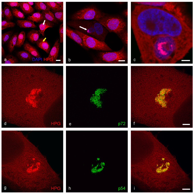
Incorporation of HPG into proteins in Vero cells. Cells were incubated with HPG for thirty minutes after forty-five minutes starvation in methionine free media, then fixed. Cells were subject to Click-IT reaction with azide conjugated to Alexa 555. Confocal image of uninfected (a) and 16 hpi (b,c) cells show an accumulation of nascent protein in cellular organelles (a, yellow arrow) and nucleoli (a, white arrow) and in the viral factory in infected cells (b, white arrow and c). By STED microscopy (d–i), the HPG labelling co-localized with both p72 (17LD3) and p54 (RB7) labelling. Scale bars: a, b = 10 µm, c, f, i = 3 µm.

