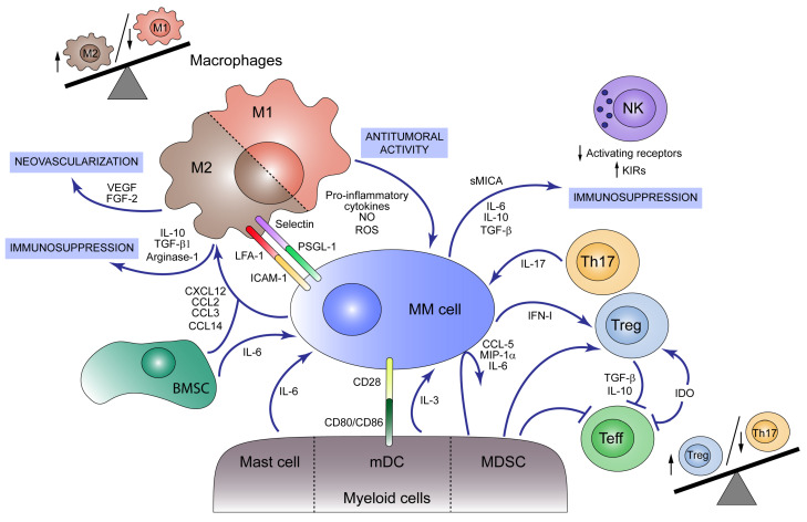
Figure 2.
Immunosuppressive tumor microenvironment (TME) in multiple myeloma (MM). Both myeloid and lymphoid cells present in bone marrow (BM) participate in MM development. M1 macrophages act as antitumoral agents producing pro-inflammatory cytokines, nitric oxide (NO) and reactive oxygen species (ROS) and acting as antigen-presenting cells (APC) that activate immune response against MM cells. MM cells and BM mesenchymal stromal cells (BMSCs) produce the chemokines CXCL12, CCL2, CCL3, and CCL14, which promote macrophage M2 polarization and migration to the tumor niche, unbalancing M1/M2 ratio towards the M2 population. M2 macrophages play a pro-tumoral role secreting immunosuppressive agents such as IL-10, TGF-β1 and Arginase-1 and neovascularization factors like VEGF and FGF-2. M2 macrophages act as MM cell protectors through the expression of selectins and LFA-1 in the macrophage surface, which binds to PSGL-1 and ICAM-1 in the plasma cell, respectively. Mast cells, although contributing to an initial tumoricidal host response, secrete IL-6 which promotes MM cell growth. Myeloid dendritic cells (mDCs) foster MM cell proliferation and survival by cell–cell interactions with tumor cells via CD80/CD86-CD28 and secretion of IL-3. Additionally, myeloid-derived suppressor cells (MDSCs) induce the secretion of cytokines such as CCL5, MIP-1α and IL-6 by MM cells and modulate the cytotoxic T cell responses inhibiting effector T cell functions and activating regulatory T cells (Treg). Other signals that promote Treg cell expansion are IFN-I, produced by MM cells, and indoleamine 2, 3-dioxygenase (IDO) activity which at the same time inhibits effector T cells. Cytokines produced by Tregs, such as IL-10 and TGF-β, together with other MM cell-derived signals attenuate effector T cell function. Th17 lymphocytes are also present in BM from MM patients and foster tumor growth through IL-17 secretion. NK cell cytotoxic activity is modulated during disease progression by cytokines and soluble factors (e.g., sMICA) secreted by MM cells that induce a reduction in activating receptors and an increase in inhibitory receptors (KIRs).

