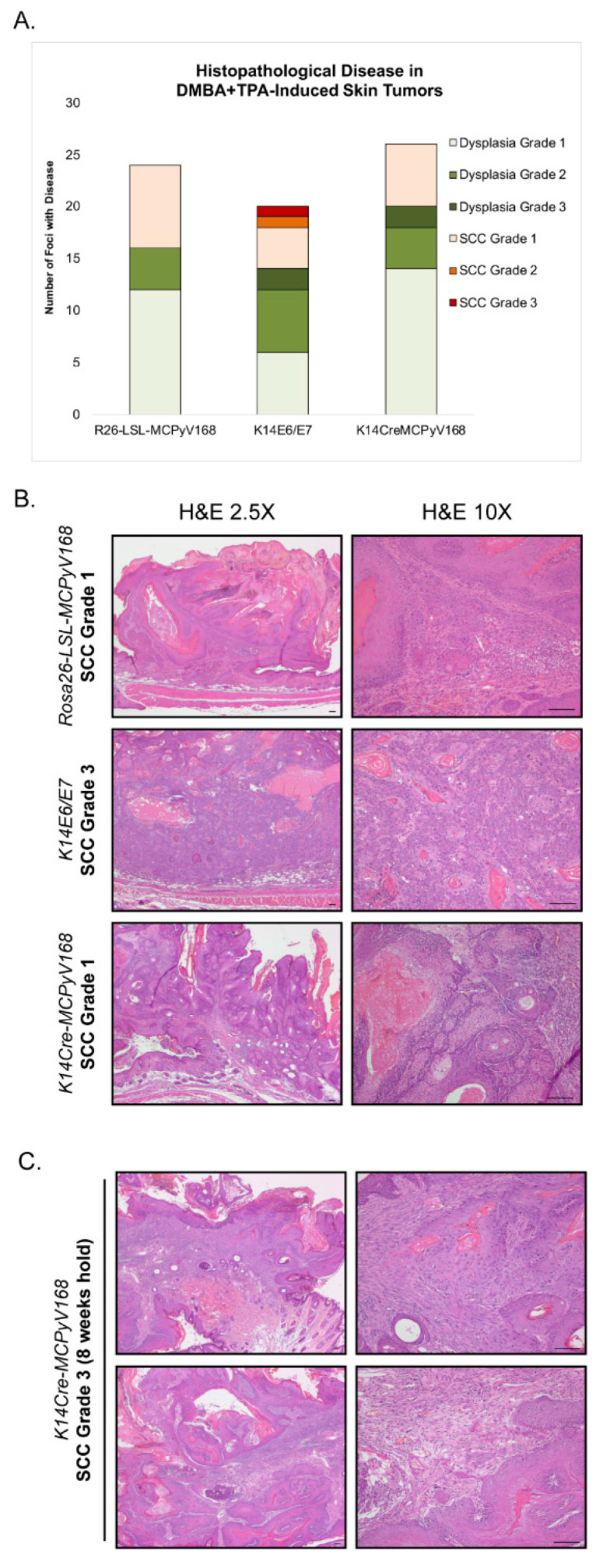
Figure 3.
Assessment of malignant progression. (A) After the 20 week DMBA+TPA treatment period, tumor-bearing Rosa26-LSL-MCPyV168, K14E6/E, and K14Cre-MCPyV168 mice were held without further treatment for an additional 5 weeks. Skin was harvested, sectioned into 5 μM sections and placed on glass slides, and stained with hematoxylin and eosin (H&E). Tissues were evaluated for histopathological disease. Each foci/tumor evaluated was given a score for worst disease among the following grades: Dysplasia Grade 1, Dysplasia Grade 2, Dysplasia Grade 3, Squamous Cell Carcinoma (SCC) Grade 1, SCC Grade 2, or SCC Grade 3. The number of foci with each disease score in each group of mice is indicated in the bar graph. The overall disease severity between groups was not statistically significant (two-sided Wilcoxon rank-sum test; all p-values > 0.17 for all comparisons). (B) Representative H&E-stained images of tissue sections from tumors harvested from DMBA+TPA-treated Rosa26-LSL-MCPyV168, K14E6/E7, and K14Cre-MCPyV168 mice after 5 weeks hold. Images show representative examples of worst disease state present in each treatment group. All scale bars = 100 μM. (C) Representative H&E-stained images of tissue sections from tumors harvested from a DMBA+TPA-treated K14Cre-MCPyV168 mouse after 8 weeks hold. Images show representative examples of worst disease state present. All scale bars = 100 μM.

