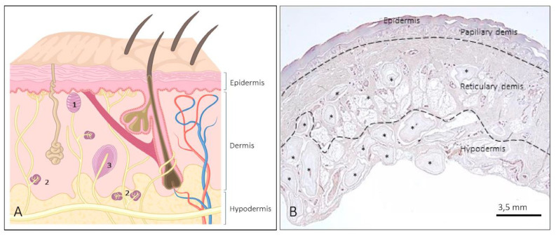
Figure 1.
(A) Schematic representation showing the structure of the skin and the different morphotypes of sensory corpuscles. 1: Meissner corpuscles; 2: Ruffini corpuscles; 3: Pacini corpuscles; (B) low magnification of a histological section of human digital skin. The lines indicate the approximate limits of the three skin layers (epidermis, dermis—papillary and reticular—and hypodermis). Both the dermis and hypodermis contain abundant sensory corpuscles. * denotes Pacinian corpuscles.

