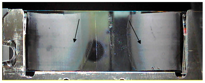
Figure 1.
Two-microliter blood drops smeared on two ends of the same indium tin oxide (ITO) slide, with the arrows pointing to optimal cell dispersion zones: cells lying close to each other disable unequivocal mass spectrometry imaging (MSI) analysis, while too much dispersion of cells yields too few cells.

