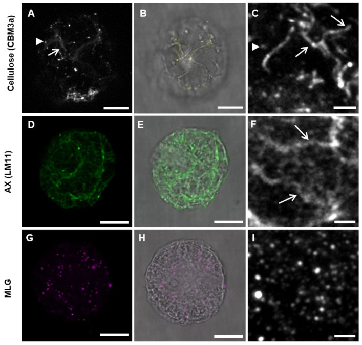
Figure 7.
Lolium SCC protoplasts treated with 200 nM isoxaben for 24 h prior to fixation and antibody labeling. (A,B,D,E,G,H) whole cell images. (C,F,I) are higher magnification images of cell wall labeling. The pattern of labeling of crystalline cellulose with CBM3a (A–C) appeared to be disrupted with possibly terminated filaments (arrows) and only partial coverage of cellulose labeling over the cell. Punctate dots were present (arrow heads). Labeling of AX using LM11 (D–F) was less disrupted. Filamentous-like structures of AX (arrows) were still present and the amount of labeling appeared to be similar to untreated cells. The punctate pattern of MLG labeling (G–I) looked similar to cells with no treatment. Scale bars: A–B, D–E, and G–H = 5 μm, and C, F, and I = 2 μm.

