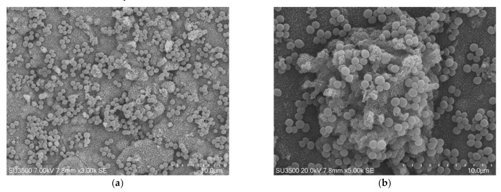
Figure 1.
S. mutans prepared by conventional scanning electron microscope (SEM) protocol. (a) SEM, 3000×. S. mutans spherical bacterial cells were scattered on biofilm matrix compact surface. Image was captured from the same sample observed in Figure 1a from [50]; (b) SEM, 5000×. Increasing magnification S. mutans spherical bacterial cells appeared clustered in small groups on the surface of a rough and dense extracellular matrix (Eps).

