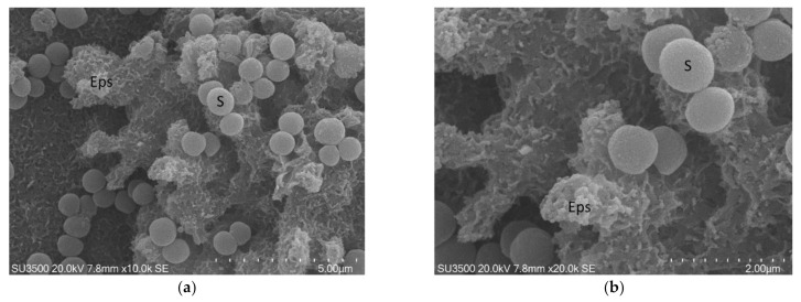
Figure 2.
S. mutans prepared by conventional SEM procedure. (a) SEM, 10,000×. Eps forms a canalicular system of compact trabeculae with spiny surface. Bacterial cells, S, are adherent to the Eps surface. Eps: extracellular polymeric substance. Image was captured from the same sample observed in [50] Figure 1d; (b) SEM, 20,000×. Bacterial cells appear irregular, Eps micro-canalicular system is not developed, only superficial holes are visible. Bacterial cells lay down on the Eps surface, and they appear naked, without a matrix covering. Bacterial cells are sometimes fragmented or indented; Eps showed a compact aspect due to the collapse of its fine structure. Bacterial cells, uncovered by the matrix, rest on the Eps surface. Eps: extracellular polymeric substance, S: S. mutans. Image was captured from the same sample observed in [50]. Figure 1f.

