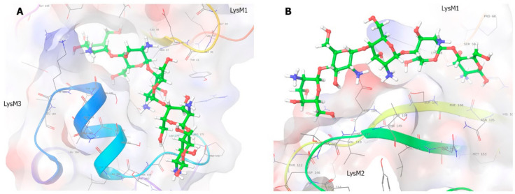
Figure 6.
Molecular modeling of PsLYK9-ECD binding with CO5. (A) Binding site 1: This figure shows the computed bonds between PsLYK9-ECD and CO5 at site 1. CO5 binds PsLYK9-ECD at site 1 along the groove above the LysM3 domain and partly in the cavity between LysM1 and LysM3. (B) Binding site 2: The figure shows the complex-forming bonds at site 2 for a pose with the best score. The complex at site 2 is formed by the protein and the ligand above the groove in the LysM2 and in the cavity between the LysM1 and LysM2 domains. Ligand is attached outwardly at site 2; thus, CO5 is exposed to the solvent more than at site 1.

