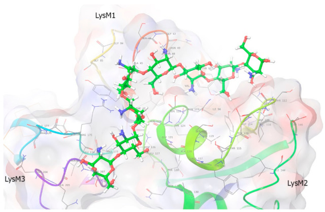
Figure 7.
Molecular modeling of PsLYK9-ECD binding with CO8-DA. The main ligand conformation pattern is partly placed at the groove between LysM1 and LisM2 (four upper ligand rings), approaching LysM2, whereas the rest of the ligand descends to the central groove between LysM3 and LysM2. The type of binding is partly hydrophobic (at the area of LysM2 domain) and partly polar at the side of binding between LysM2 and LysM3. The lower and upper CO8-DA molecular rings are half-available to the solvent.

