Figure 3.
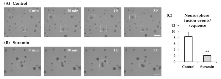
Effect of suramin treatment on neurosphere fusion. Neural progenitor cells of the postnatal rat subventricular zone were cultured for 72 h in the absence (control) or presence of 200 μM suramin. Time-lapse imaging was performed after 48 h of seeding over 21 h of culture in 10-min intervals. (A) Images captured from a time-lapse sequence showing neurospheres in control conditions. Dashed squares illustrate two examples of adjacent neurospheres that undergo adhesion (see t = 20 min) and fusion (t = 3 and 4 h). Bar: 200 μm. (B) Images captured from a time-lapse sequence showing neurospheres in a culture treated with 200 μM suramin. Dashed squares illustrate an example of adjacent neurospheres that undergo adhesion (see t = 20 min) and fusion (t = 3 and 4h). Bar: 200 μm. (C) Quantification of neurosphere fusion events per time-lapse sequence in each experimental condition (control or suramin). Data are mean ± SEM (n = 8, ** p < 0.01; Student’s t-test).

