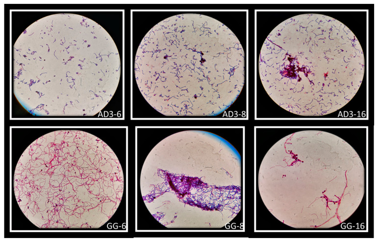
Figure 7.
Macroscopically visible clumps after Giemsa staining of L. rhamnosus AD3 and GG with all three Candida strains. Co-aggregates of L. rhamnosus AD3 with C. albicans (AD3-6), C. krusei (AD3-8) and C. tropicalis (AD3-16). Co-aggregates of L. rhamnosus GG with C. albicans (GG-6), C. krusei (GG-8) and C. tropicalis (GG-16).

