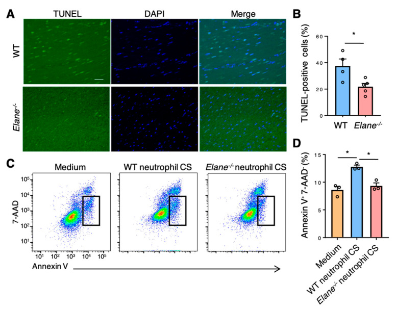
Figure 5.
NE deficiency reduced cardiomyocyte apoptosis in vivo and in vitro. (A) Representative TUNEL staining images of WT and Elane−/− heart sections on day 1 post-MI. Scale bar = 50 μm. (B) The bar graph shows the TUNEL-positive cells as a percentage of DAPI-positive cells in WT and Elane−/− hearts on day 1 post-MI. Results are presented as mean ± SEM, n = 5, * p < 0.05. (C) Representative flow cytometric plots showing Annexin V+ and 7-AAD− apoptotic HL-1 cells. HL-1 cells were cultured in the CS of WT or Elane−/− neutrophils. Results are presented as mean ± SEM, n = 3 each, * p < 0.05. CS, culture supernatant.

