Figure 1.
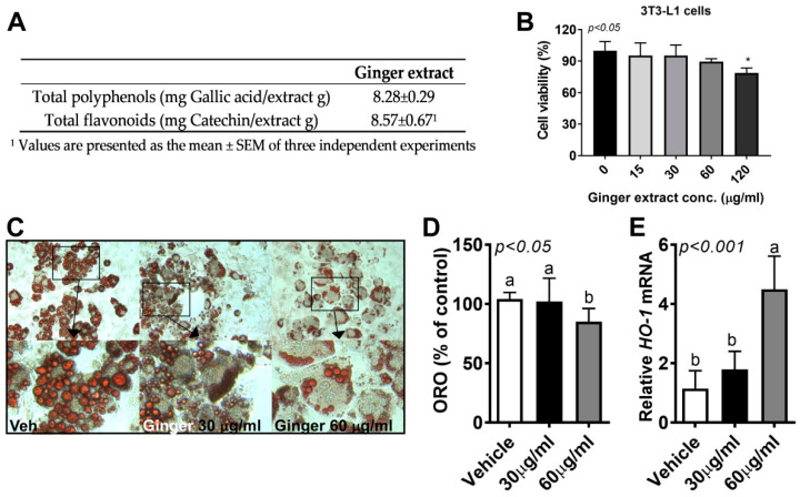
Ginger reduced lipid accumulation in 3T3-L1 adipocytes. (A) Total polyphenol and flavonoid contents of ginger extract; the culture of 3T3-L1 cells was incubated with ginger extract at different doses (15–120 μg/mL). 2,3-Bis-(2-methoxy-4-nitro- 5-sulfophenyl)-2H-tetrazolium-5-carboxanilide salt (XTT) reagent was added for 3 h into 96-well plates to measure cell viability. (B) Cell viability by XTT; the 3T3-L1 cells were differentiated in the presence or absence of ginger extract (0–60 μg/mL) for 7 days. Dimethyl sulfoxide (DMSO) was used as a vehicle control. (C) Triglyceride (TG) accumulation in 3T3-L1 adipocytes visualized using Oil Red O (ORO) staining. Representative images from three separate experiments are shown (20× magnification, upper). Images were cropped to the relevant part of the field without altering the resolution (close-cropped images, lower). (D) ORO dye was extracted using isopropanol and quantified as the optical density at 500 nm (OD500). (E) Heme oxygenase 1 (HO-1) gene expression determined using real-time PCR. Data are represented as the mean ± standard error of the mean (SEM) of three independent experiments. * p < 0.05 (compared with vehicle). Bars with different letters are significantly different according to one-way ANOVA with Bonferroni’s comparison test.

