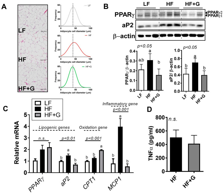
Figure 4.
Ginger supplementation attenuated HF-induced adipocyte hypertrophy. Male 6-week-old C57BL/6 mice were fed an LF (white), HF (black), or HF + G (gray) diet for 7 weeks (n = 4–5 per group). (A) H&E staining of adipose tissue (20× magnification) and adipocyte size distribution (Gaussian curve fitting). (B) protein expression of peroxisome proliferator-activated receptor γ (PPARγ) and adipocyte protein 2 (aP2) in epididymal adipose tissue, quantified by Western blot. β-Actin was used as a loading control. (C) mRNA expression of PPARγ, aP2, CPT1, and MCP1 in epididymal adipose tissue determined using real-time PCR. (D) TNFα cytokine production by ELISA. Data are expressed as the mean ± SEM (n = 4–5). n.s. represents no significance. Bars with different letters are significantly different according to one-way ANOVA with Bonferroni’s comparison test.

