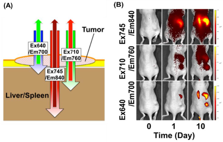
Figure 3.
Depth-dependent NIR fluorescence in vivo imaging of a tumor. (A) A schematic illustration of depth-dependent NIR fluorescence imaging. Longer excitation and emission wavelengths, such as 745/840 nm (Ex745/Em850), can penetrate tissue very well and can detect particles in deeper sites, compared to shorter wavelengths (i.e., Ex710/Em760 and Ex640/Em700). (B) We administered totals of 2 mg (Day 1) and 6 mg (Day 10) of thiol-OS/IR820 into a mouse that had a subcutaneous xenograft tumor intravenously and observed the fluorescence using the in vivo imaging system under three excitation/emission wavelength conditions (Ex640/Em700 nm, Ex710/Em760 nm, and Ex745/Em840 nm). Reproduced with permission from Nakamura, M.; Hayashi, K.; Nakamura, J.; Mochizuki, C.; Murakami, T.; Miki, H.; Ozaki, S.; Abe, M. Near-Infrared Fluorescent Thiol-Organosilica Nanoparticles That are Functionalized with IR-820 and Their Applications for Long-Term Imaging of in Situ Labeled Cells and Depth-Dependent Tumor in Vivo Imaging. Chem. Mater. 2020, 32, 7201–7214 [106].

