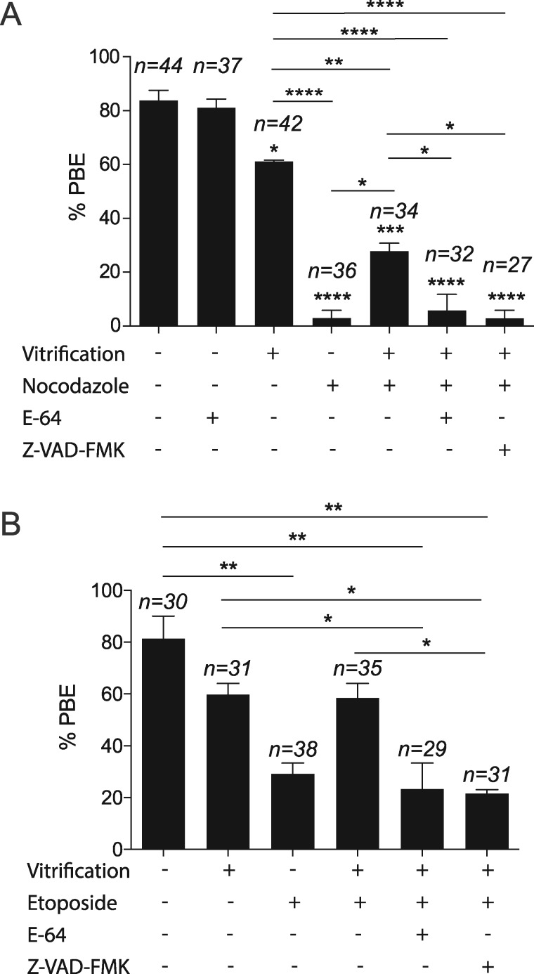
Figure 3.
Effect of mouse oocyte vitrification on PBE. Fresh and vitrified/thawed GV oocytes were treated with nocodazole (A) or etoposide (B) to maintain the SAC active, and divided into the indicated treatment groups (dimethylsulphoxde (DMSO), E-64 or Z-VAD-FMK), in vitro matured for 16 h and scored to quantify the percentage extrusion of the first polar body (PBE). The total number of analyzed oocytes in each group (from two independent replicates) is specified above each condition within each graph. The data are expressed as mean ± SEM; one-way ANOVA was used to analyze the data. Values with asterisks vary significantly, *P < 0.05, **P < 0.01, ***P < 0.001, ****P < 0.0001. Unless otherwise specified, asterisks above each group denote the statistical analysis of P-value against the control group.

