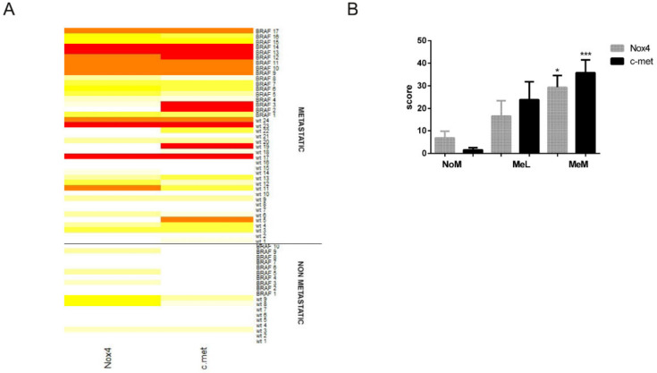
Figure 1.
Nox4 and c-met expression in primary melanoma. (A) Heat-map showing the Nox4 and c-met IHC staining in primary melanoma, NoM and MeM, WT or with BRAF mutation. White means 0% of positive cells, intense red means 100%, yellow and orange mean levels in between. (B) Graph showing immunohistochemical (IHC) evaluation, dividing metastatic melanoma in MeL (positivity only in lymph nodes) and MeM (distant metastasis). * p-value < 0.05; *** p-value < 0.001.

