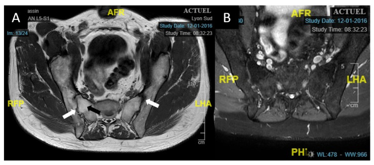
Figure 2.
Axial sacroiliac MRI of case 1. (A): T1-weighted sacroiliac axial MRI of case 1 showing major joint reshaping in the form of hypo dense areas bordering the joint (white arrows) as well as ankylosis of the right sacroiliac joint with a T1 hypersignal of the subchondral bone. (B): T2 SPAIR axial sequence without hypersignal of the sacroiliac joints, indicating the absence of active involvement. Abbreviations: A = anterior, P= posterior, R = right, L = left, H = head, F = foot; MRI: magnetic resonance imaging; T2 SPAIR: T2 Spectral Attenuated Inversion Recovery.

