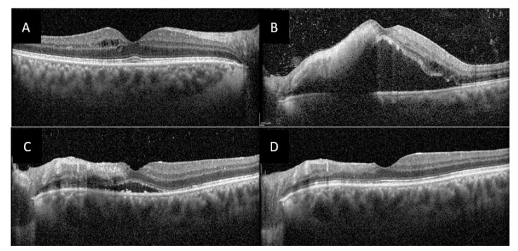
Figure 4.
Optical coherence tomography (OCT) of both macula of case 2 patient. (A), OCT of the right macula showing a few cysts of intraretinal edema. (B), OCT of the left macula showing a large serous retinal detachment, a hyperreflective nodule in the inner retinal layers, intraretinal edema and numerous hyperreflective dots in the vitreous. (C), OCT of the left macula at 1-month follow-up showing partial regression of subretinal fluid. (D) OCT of the left macula at 3 months follow-up showing complete regression of macular edema, and atrophy of internal retina in place of the hyperreflective nodule.

