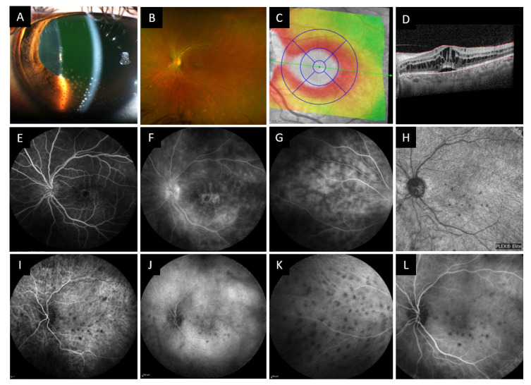
Figure 7.
Slit lamp examination, wide field fundus photography, OCT, fluorescein angiography and OCT angiography of case 3 patient. (A) Slit lamp examination showing numerous mutton fat keratic precipitates with anterior chamber reaction and posterior synechiae. (B) Wide field fundus photography showing macular edema and inferior yellow-white waxy spots. (C) OCT Thickness mapping of the macula and B-scan (D) showing cystoid macular edema with retro foveolar subretinal detachment. (E–G) Fluorescein angiography showing signs of cystoid macular edema, papillitis, capillaritis and choroidal granulomas. (I–K) Indocyanine angiography showing hypofluorescent spots most visible in the early phase, present at the posterior pole and periphery. (H) OCT angiography of the choriocapillaris at diagnosis showing spots of reduced choriocapillary flow corresponding to choroidal granulomas on the ICG angiogram (L).

