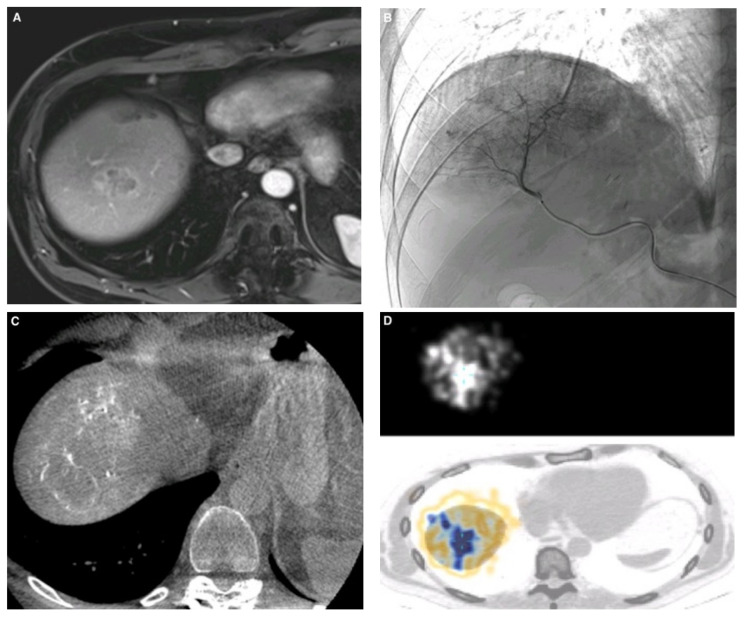
Figure 1.
51-year-old man with a single liver metastasis from colorectal cancer. (A) Hepatic magnetic resonance imaging (MRI) before Y90 transarterial radioembolization (TARE) showing contrast enhancement of the metastasis in the hepatic dome. (B) Selective angiography of arteries supplying the VII and VIII segments of the liver, obtained with a Progreat 2.4 microcatheter before the injection of Y90 microparticles. (C) Cone beam CT with contrast injection showing good targeted-lesion coverage with the microcatheter in the appropriate position. (D) Photon-emission tomography (PET) CT scan performed on the day following TARE, showing uptake by the targeted liver tumor.

