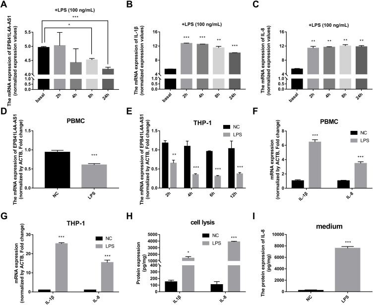
Figure 2.
Expression of EPB41L4A-AS1 and inflammatory factors in PBMC and THP-1 cells induced by LPS. (A–C) Expression of EPB41L4A-AS1 and inflammatory factors IL-1β and IL-8 at different times (basal, 2, 4, 8, and 24 h) during LPS treatment of PBMC (GEO dataset GDS2856). (D) EPB41L4A-AS1 expression in PBMC after 1 μg/mL LPS treatment for 24 h (n=3). (E) EPB41L4A-AS1 expression in THP-1 cells after 1 μg/mL LPS treatment for 2–12 h (n=3). (F) IL-1β and IL-8 expression in PBMC after 1 μg/mL LPS treatment for 24 h (n=3). (G) IL-1β and IL-8 expression in THP-1 cells after 1 μg/mL LPS treatment for 6 h (n=3). (H) IL-1β and IL-8 protein expression in cell lysate of THP-1 cells after 1 μg/mL LPS treatment for 6 h (n=3). (I) IL-8 protein expression in medium of THP-1 cells after 1 μg/mL LPS treatment for 6 h (n=3). NC indicates negative control. Data are shown as mean ± SD. *P<0.05, **P<0.01, ***P<0.001; Student’s t-test.

