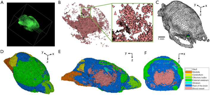
Figure 1.
Digital mouse brain model with blood vasculature incorporated for 3D Monte Carlo simulation. The blood vasculature information was obtained from light sheet fluorescence microscopy (LSFM) imaging. The LSFM image was scaled and shifted manually to register with Digimouse model. The merged model was then converted to tetrahedral mesh. (A) 3D rendering of LSFM blood vasculature image. (B) Blood vasculature distribution in the model, assuming whole blood with a 45% hematocrit. The thalamus and cortex have a relatively high vessel distribution density. (C) Skull in the Digimouse model. (D) Overview of the merged model. Different colors represent different regions identified in brain. Skin and skull are not shown. (E) A representative sagittal plane around the middle of the brain. (F) A representative coronal plane around the bregma. Digimouse model displayed here used a much larger element size for better visualization of the structure.

