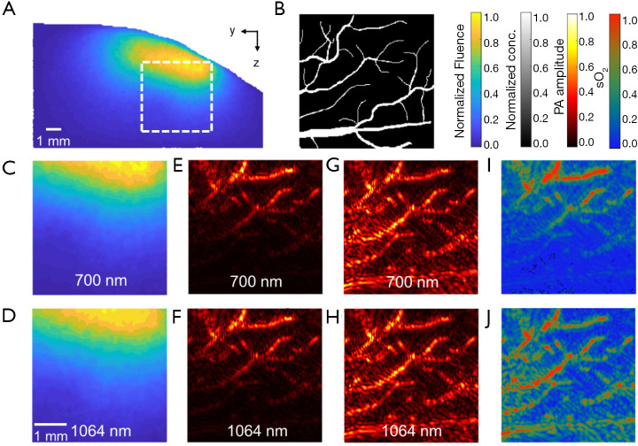
Figure 9.
Simulated photoacoustic image reconstruction using k-Wave. Blood oxygenation was calculated using linear spectral unmixing. (A) 700 nm fluence map at the imaging plane (x=6 mm) in normalized absolute scale, corresponding to a FOV of 7×12.8 mm2. A 4×4 mm2 subregion (white dashed box) was segmented out for detail examination. (B) Ground truth hemoglobin concentration map. The vessel is assumed to have a blood oxygenation level of 85%. (C,D) Normalized fluence map in the subregion at 700 and 1,064 nm, respectively. (E,F) Reconstructed images without correcting the fluence at 700 and 1,064 nm, respectively. (G,H) Reconstructed images corrected by the fluence map at 700 and 1,064 nm, respectively. (I,J) Estimated blood oxygenation without (E,F) and with (G,H) fluence correction, respectively.

