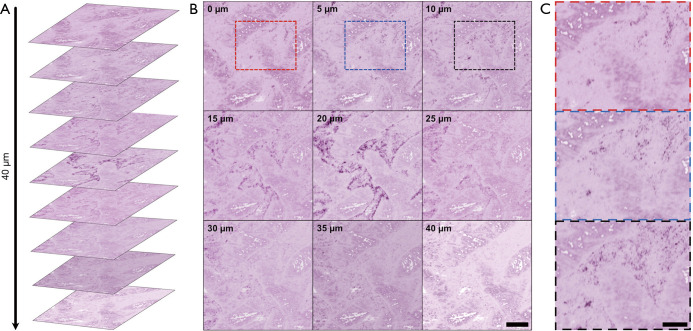
Figure 3.
Optically sectioned PARS volumetric images within formalin fixed paraffin embedded (FFPE) human gastrointestinal tissues. (A) Optical sections of differing depths in FFPE human gastrointestinal tissues arranged in the same orientation as imaging; (B) optical sections of differing depths in FFPE human gastrointestinal tissues. Scale bar: 200 µm; (C) notable areas of structural changes between the 0 to 10 micron depth are enclosed in the red blue and black boxes. Scale bar: 100 µm. Artificial H&E-like coloring has been applied to the PARS images.

