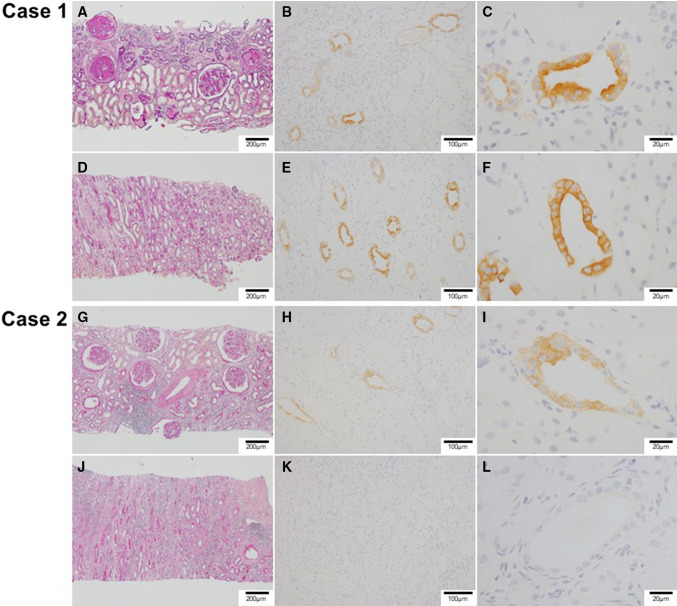
Fig. 3.
Microscopic findings of kidney samples in responder (case 1 [a–f]) and non-responder (case 2 [g–l]). a Case 1: severe glomerular sclerosis, indicating diabetic nephropathy, and severe tubular atrophy in the renal cortex. PAS (Periodic Acid-Schiff) stain. b Case 1: immunohistrochemical staining showing expression of aquapolin-2 in the collecting duct of the renal cortex. c Case 1: high-power filed of the renal cortex showing stronger staining of aquapolin-2 in the lumen side. d Case 1: relatively preserved structures of tubules in the renal medulla. PAS stain. e Case 1: immunohistrochemical staining showing expression of aquapolin-2 in the medullary collecting duct. f Case 1: high-power filed of the renal medulla showing stronger staining of aquapolin-2 in the lumen side. g Case 2: slight tubular atrophy and interstitial inflammatory cell infiltration in the renal cortex. Relatively preserved structures of glomeruli, indicating diabetic nephropathy. PAS stain. h Case 2: immunohistrochemical staining showing weak expression of aquapolin-2 in the renal cortex collecting duct. i Case 2: high-power field of renal cortex showing weak staining of aquapolin-2. j Case 2: severe interstitial inflammatory cell infiltration and tubular atrophy in the renal medulla. PAS stain. k Case 2: aquapolin-2 immunostaining was negative in the renal medulla. l Case 2: high-power field of renal medulla showing no staining of aquapolin-2

