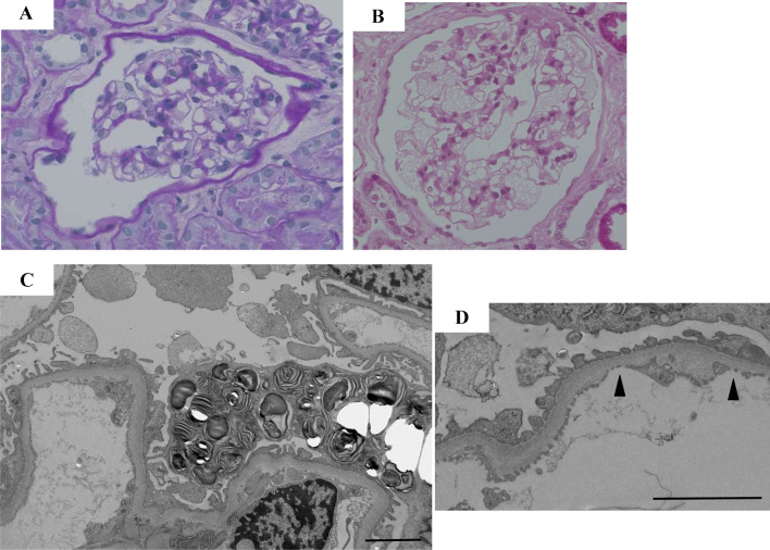
Fig. 1.
a, c, d Renal pathology prior to kidney donation; b a 1-h biopsy. a No apparent change was detected in the glomeruli under a light microscopic examination before kidney donation (PAS staining × 400). b A 1-h biopsy revealed remarkable swelling and obvious vacuolation of the glomerular podocytes (PAS staining × 400). c Pre-donation electron microscopy revealed numerous zebra bodies in the podocytes (× 3000). d Basement membrane thinning (180 nm) was observed in part of the specimen (Arrowhead). Scale bars 2 μm

