Fig. 5.
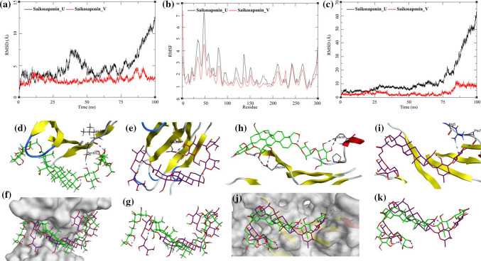
Trajectory analysis for human interleukin-6 (PDB: 1N26) bound to Saikosaponin_U and Saikosaponin_V; a root-mean-square deviation (RMSD), b root-mean-square fluctuations per amino acid (aa) (RMSF) and c root-mean-square deviation for each ligand (Lig-RMSD). Interaction analysis of the human interleukin-6 (PDB: 1N26) bound to Saikosaponin_U during the molecular dynamics simulation; d equilibrated structure of Saikosaponin_U (green) bound to the human interleukin-6 before MDS production phase; e Saikosaponin_U (purple) bound to the human interleukin-6 post-MDS production phase; f receptor surface analysis; g ligand analysis. Interaction analysis of the human interleukin-6 (PDB: 1N26) bound to Saikosaponin_V during the molecular dynamics simulation; h equilibrated structure of Saikosaponin_V (green) bound to the human interleukin-6 before MDS production phase; i Saikosaponin_V (purple) bound to the human interleukin-6 post-MDS production phase; j receptor surface analysis; k ligand analysis

