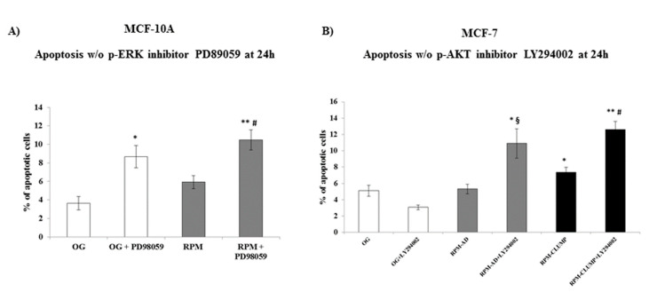
Figure 12.
Apoptosis analysis in MCF-10A (A) and MCF-7 (B) cells at 24 h after exposition to weightlessness w/o the p-ERK inhibitor, PD89059, and w/o the p-AKT inhibitor, LY294002, respectively. Bar charts/graphs show the percentage of apoptotic cells (Annexin V+/7-AAD-); each column represents the mean value ± SD of three independent experiments. In MCF-10A, * p < 0.05, ** p < 0.01 vs. OG; # p < 0.05 vs. RPM by ANOVA followed Bonferroni post-test. In MCF-7 * p < 0.05, ** p < 0.01 vs. OG; § p < 0.05 vs. RPM-AD; # p < 0.05 vs. RPM-CLUMP by ANOVA followed by Bonferroni post-test.

