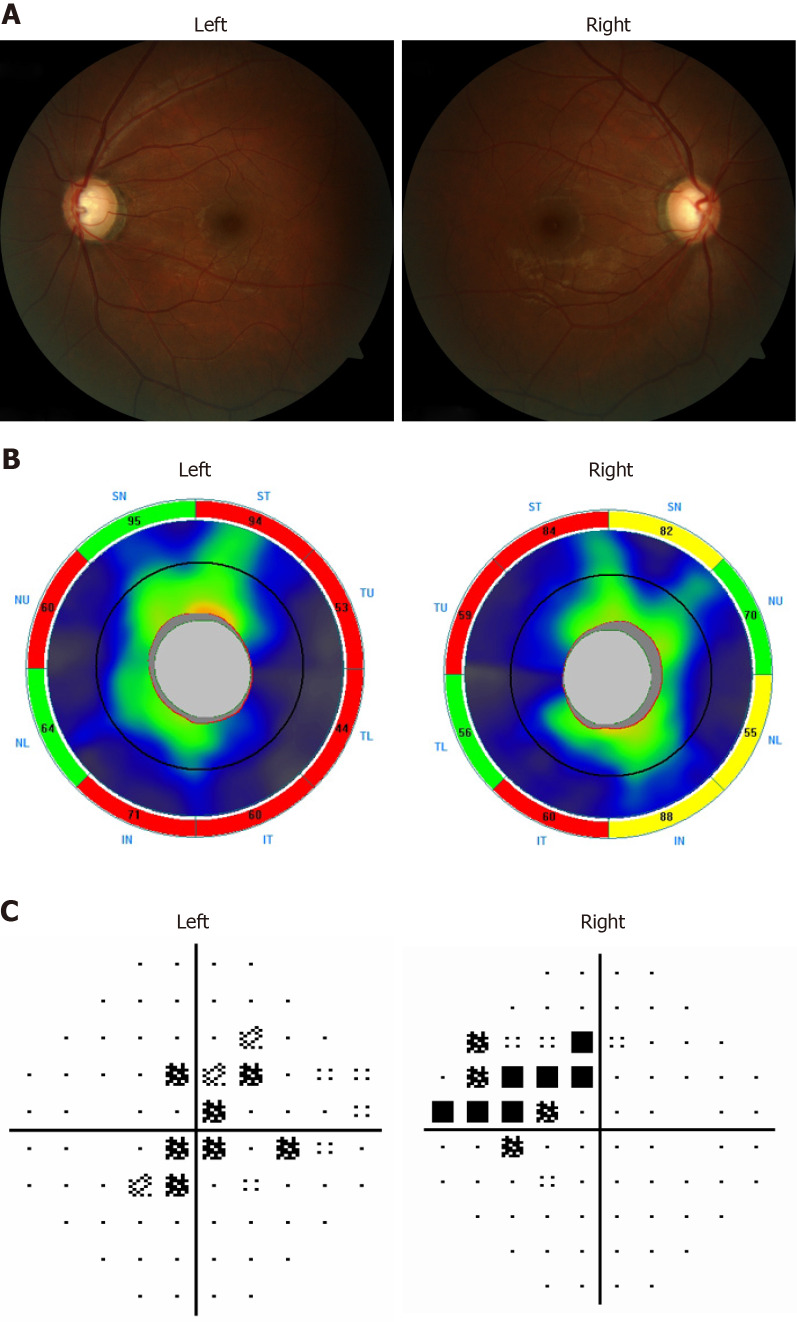
Figure 1.
Ocular assessments of the proband. A: Fundus photo of the right and left eye; B: Ocular coherence tomography-retinal nerve fiber layer of both eyes (Key: S: Superior; T: Temporal; N: Nasal; I: Inferior); C: Right and left nasal step visual field defects consistent with glaucomatous damage.

