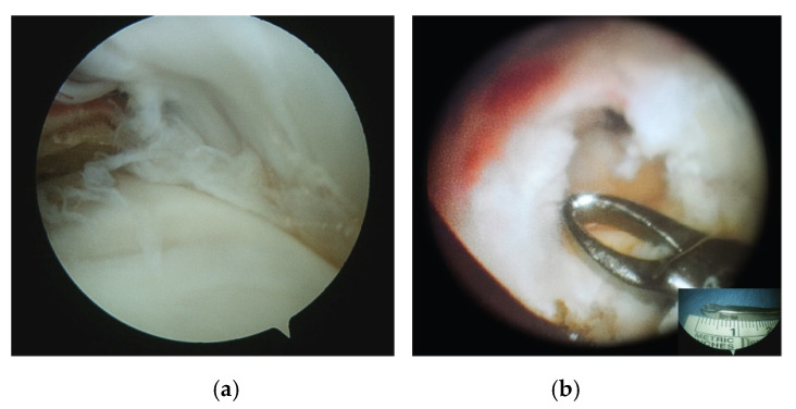
Figure 2.
(a) Arthroscopic view of an endoarticular tear of the ST (posterior portal). (b) Arthroscopic view of the bursal surface of ST after the removal of the “critical zone,” a small area of decreased vascularity about 10 mm from the proximal insertion of the ST (lateral portal). Insert shows the size of the forceps’ edge (about 10 mm); ST, supraspinatus tendon.

