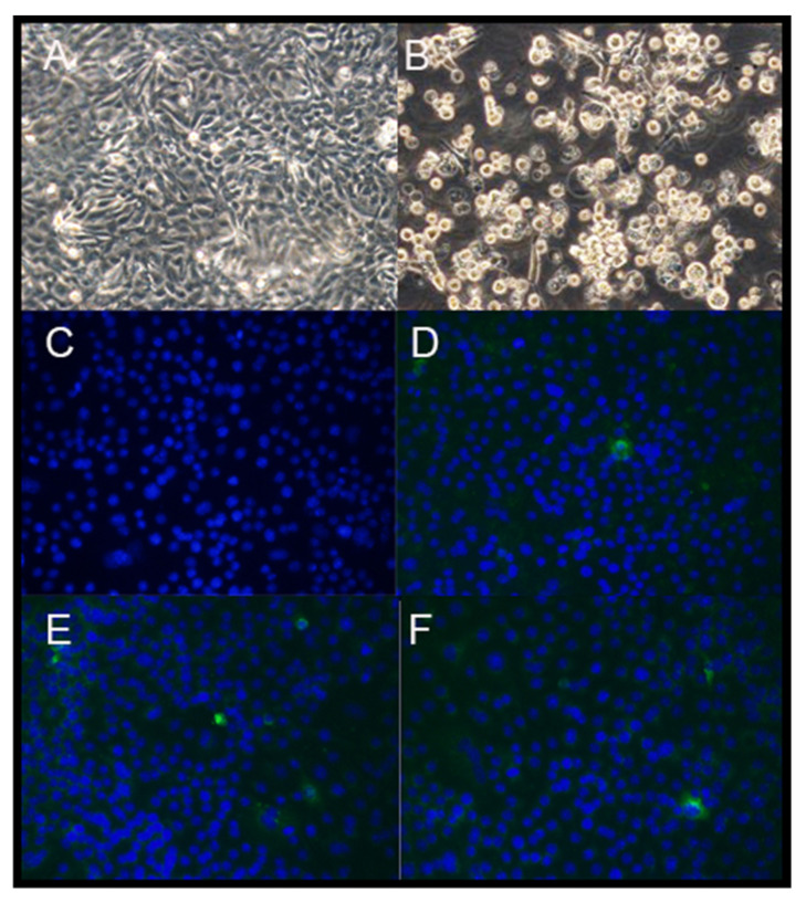
Figure 2.
The rescue of the recombinant PDCoVs (magnification, 100×). LLC-PK1 cells transfected with N transcript only (A) and the full-length mRNA genome and N transcript of PDCoV (B) at 72 h post electroporation (hpi) viewed by light microscopy. IF staining for cells inoculated with control (C), icPDCoV (D), icPDCoV-RBDISU (E), and icPDCoV-SHKU17 (F) at 24 hpi [PDCoV N gene (in green) and nuclei (in blue)].

