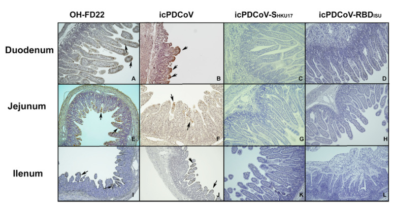
Figure 6.
Immunohistochemistry staining of PDCoV N proteins in the enterocytes of duodenum, jejunum, and ileum of piglets inoculated with OH-FD22 (A,E,I), icPDCoV (B,F,J), icPDCoV-SHKU17 (C,G,K), or icPDCoV-RBDISU (D,H,L) (magnification, 100×). The brown signals, indicated by arrows, represent the PDCoV antigens in enterocytes and were observed in the OH-FD22- and icPDCoV-inoculated, but not in icPDCoV-SHKU17- or icPDCoV-RBDISU-inoculated and mock piglets (data not shown).

