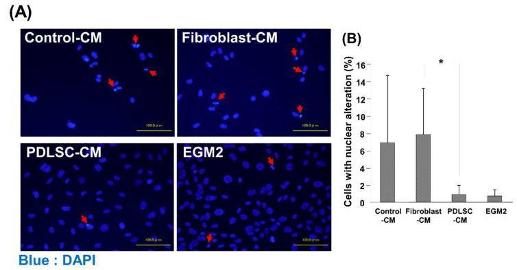
Figure 3.
Cell death of HUVECs cultured in various CMs. Representative images of HUVECs with alterations in nuclear morphology after DAPI staining are shown, including nuclei fragmentation and condensation (A). The number of HUVECs with nuclear alteration was quantified (B). A lower number of cells with alterations of nuclear morphology was identified in HUVECs cultured with PDLSC-CM, compared to Control-CM and Fibroblast-CM. Red arrows: alterations of nuclear morphology (* p < 0.05) (HUVECs = human umbilical cord vein endothelial cells, DAPI = 4′,6-Diamidino-2-phenylindole dihydrochloride, CM = conditioned medium, PDLSCs = periodontal ligament stem cells, EGM2 = endothelial cell growth medium 2). Data from an independent experiment performed three times were shown. (n = 3).

