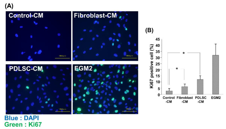
Figure 4.
Ki67 staining of HUVECs cultured in various CMs. Representative merged images of Ki67 and nucleus (DAPI) staining in HUVECs cultured in Control-CM, Fibroblast-CM, PDLSC-CM, and EGM2 (A). The number of proliferating HUVECs was examined using Ki67 immunostaining (B). Quantification of the ratio of Ki67-positive cells. An increased number of Ki67-positive cells was observed in HUVECs cultured with PDLSC-CM compared to Control-CM. HUVECs cultured in EGM2 served as positive control. (* p < 0.05) (HUVECs = human umbilical cord vein endothelial cells, DAPI = 4′,6-Diamidino-2-phenylindole dihydrochloride, CM = conditioned medium, PDLSC = periodontal ligament stem cells, EGM2 = endothelial cell growth medium 2). Data from an independent experiment performed three times were shown. (n = 3).

