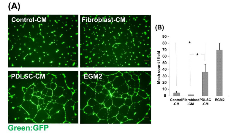
Figure 5.
Effect of CM on the network formation of HUVECs. Fluorescence microscopic images of HUVEC networks (A). HUVECs-GFP were cultured on Matrigel in the presence of Control-CM, Fibroblast-CM, PDLSC-CM, and EGM2. The number of mesh structures formed by HUVECs was quantified (B). HUVECs in PDLSC-CM formed more capillary-like networks than those in Control-CM and Fibroblast-CM. (* p < 0.05) (HUVECs = human umbilical cord vein endothelial cells, CM = conditioned medium, PDLSCs = periodontal ligament stem cells, EGM2 = endothelial cell growth medium 2). Data from an independent experiment performed three times were shown. (n = 3).

