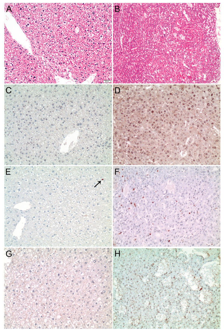
Figure 5.

MDM4 is upregulated in SRF-VP16iHep transgenic mice. (A) Normal liver parenchyma in control mice (HE staining). (B) Well-differentiated HCC in a 30-week-old SRF-VP16iHep mouse showing trabecular disarray and pseudogland formation. MDM4 immunostaining is negative in control mice (C), while a diffuse, predominantly nuclear staining is seen in SRF-VP16iHep mice (D). Individual hepatocyte nuclei (arrow) are positive for phosphorylated-ELK1 in the control liver (E), whereas the number of p-ELK1 positive nuclei is significantly increased in SRF-VP16iHep mice (F). There is no ELK4 immunosignal in control mice (G). In contrast, the SRF-VP16-induced HCC reveals weak to moderate nuclear ELK4 staining (H). Scale bar: 20 µM.
