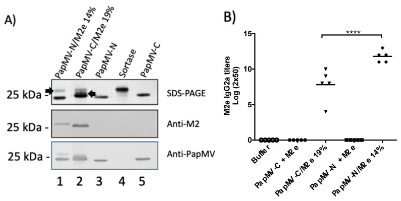
Figure 3.
Coupling of the M2e peptide to the PapMV vaccine platforms and assessment of the humoral response directed to M2e in mice. (A) The M2e antigen was coupled to the PapMV-N and PapMV-C vaccine platforms using the sortase A (SrtA) enzyme. Coupled bands for both platforms can be visualized on SDS-PAGE slightly over the 25 kDa marker. The arrow pointing to the right indicates the signal of the PapMV-N coupled to the M2e peptide (lane 1), and the arrow pointing to the left indicates the signal of the PapMV-C coupled to the M2e peptide (lane 2). Signals corresponding to PapMV-N CP (lane 3) and PapMV-C CP (lane 5) are also observed. The signal corresponding to SrtA is shown in lane 4. Remaining SrtA after the coupling reaction is seen in lanes 1 and 2. Proteins from the SDS-PAGE were transferred to a membrane to perform Western blotting using anti-M2e (middle panel) or anti-PapMV (lower panel) antibodies. (B) Balb/C mice, five per group, were immunized once, intramuscularly (i.m.), with formulation buffer (Buffer), PapMV-C (10 µg) with 1 µg of free peptide M2e, 10 µg PapMV-C/M2e 19%, PapMV-N (10 µg) with 1 µg of free peptide M2e, and 10 µg PapMV-C/M2e 14%. ELISA was performed with serum harvested at day 20 to assess immunoglobulin G (IgG) 2a titers directed to the M2e peptide. **** p > 0.0001.

