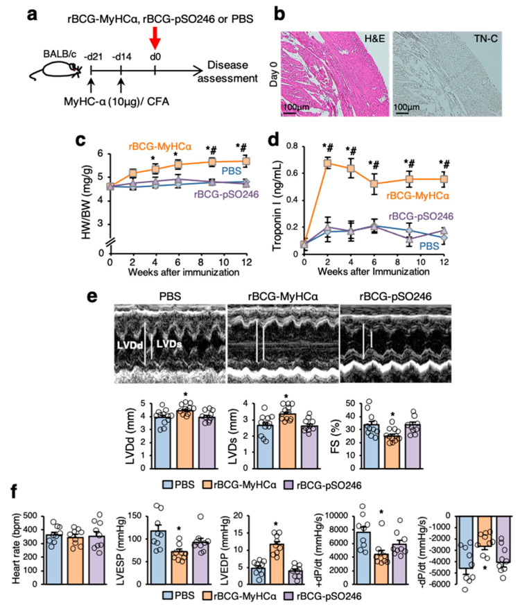
Figure 3.
rBCG-MyHCα immunization induced heart failure in mice. (a) Mice were immunized twice with 10 μg of MyHCα peptide/CFA on days 14 and 21 before rBCG immunization for priming. rBCG-MyHCα, rBCG-pSO246, or PBS were injected subcutaneously on day 0. (b) Representative histology (H&E staining and TN-C immunostaining) of the heart sections on day 0 (21 days after first immunization with MyHCα). (c,d) HW/BW (c) and serum troponin I concentrations (d) at indicated time points are shown (n = 10 each). Results are presented as mean ± SEM. * p < 0.05 vs. PBS and # p < 0.05 vs. rBCG-pSO246 by one-way ANOVA with Tukey’s post hoc test. (e) Representative M mode images of echocardiography 12 weeks after rBCG immunization. Bar graphs represent echocardiographic parameters. Results are presented as mean ± SEM, n = 9–12, * p < 0.05 vs. PBS and rBCG-pSO246 by Kruskal-Wallis analysis with a post hoc Steel-Dwass test. (f) Bar graphs represent hemodynamic parameters at 12 weeks after rBCG immunization. Results are presented as mean ± SEM, n = 9 each, * p < 0.05 vs. PBS and rBCG-pSO246 by Kruskal-Wallis analysis with a post hoc Steel-Dwass test.

