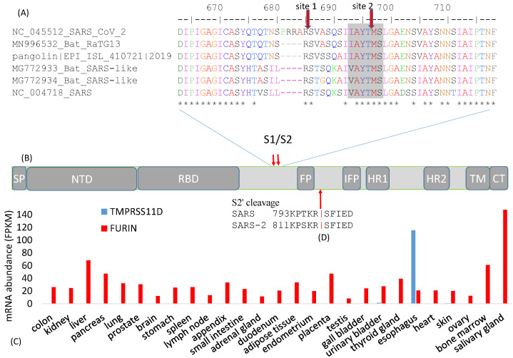
Figure 2.
Cleavage sites at the S1/S2 boundary. (A) An insertion of 12 nt in SARS-CoV-2 results in a new polybasic furin cleavage site, resulting in two cleavage sites indicated by the red downward arrows. “*” indicates sites that are identical among the six viral strains. Numbers follow (B) Schematic domain structure of S protein, with the same abbreviation as in Figure 1A; (C) Tissue-specific mRNA distribution of human trypsin-like protease TMPRESS11D and FURIN, derived from [30]; (D) Cleavage site for splitting S2 into FP and S2′ domains.

