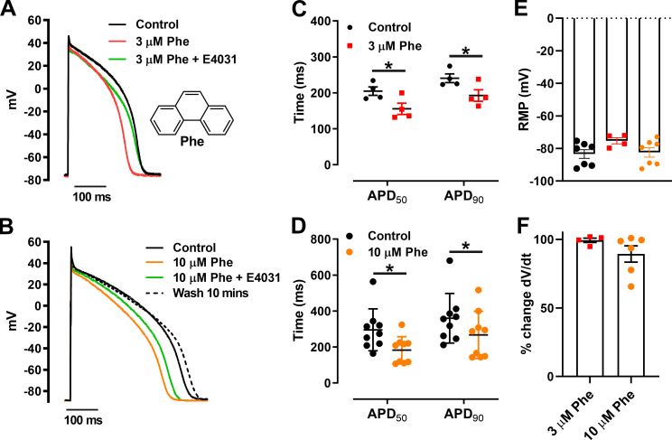
Figure 1.
Phenanthrene (Phe) shortens APD in zebrafish ventricular cardiomyocytes. (A and B) Representative AP traces at 0.5 Hz exposed to 3 µM Phe (A) and 10 µM Phe (B; with and without 2 µM E-4031 [ERG channel blocker]) with recovery from inhibition (wash). (A) Inset: Molecular structure of phenanthrene. (C and D) Scatter plot showing mean ± SEM of APD50 and APD90 in the absence and presence of 3 µM Phe (C; *, P < 0.05; Wilcoxon test) and 10 µM Phe (D; *, P < 0.05; Wilcoxon test). (E and F) No significant change in resting membrane potential (RMP; E) and dV/dt was observed in presence of phenanthrene (F). All data represented as mean ± SEM (n = 4–9; N = 5).

