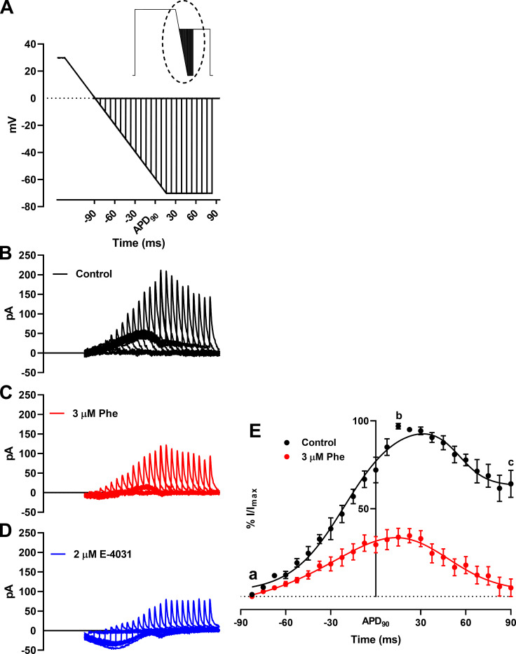
Figure 5.
Effect of phenanthrene (Phe) on response of IKr to premature stimulation. (A) Magnified view of the dotted area of (inset) paired ventricular AP-like command waveform protocol. (B–D) Representative trace of families of transient currents elicited corresponding to each depolarization in the absence (B; black) and presence (C) of 3 µM phenanthrene (red) and 2 µM E-4031 (D; blue). (E) Percentage response of peak IKr transients in the absence (black) and presence of 3 µM phenanthrene (red). Peak IKr transients for each cell under control conditions were normalized to the maximal current transient during the protocol. Peak IKr transients in the presence of 3 µM phenanthrene (red) are represented as percentage response relative to its corresponding percentage control transient. First, peak, and last transient obtained are labeled a, b, and c, respectively. All data represented as mean ± SEM (n = 7 or 8; N = 3).

