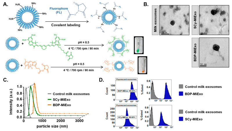
Figure 1.
Optical labeling and physicochemical characterization of control and fluorescence-labeled milk exosomes (MiExo). (A) Chemical strategies for labeling exosomes with Bodipy FL (BDP-FL, orange) and sulfo-cyanine 7.5 (SCy 7.5, green), and the resulting fluorescently labeled exosomes in dilution fluids. (B) Transmission electron microscope images for morphological evaluations of exosomes. (C) Size distributions of nanovesicles, evaluated with dynamic light scattering. Data are expressed as the mean ± standard deviation; (D) Flow cytometry results show the abundances of control exosomes (grey) and fluorescent nano-probes (blue). SCy MiExo: SCy 7.5-labeled milk exosomes; BDP-MiExo: BDP-FL-labeled milk exosomes.

