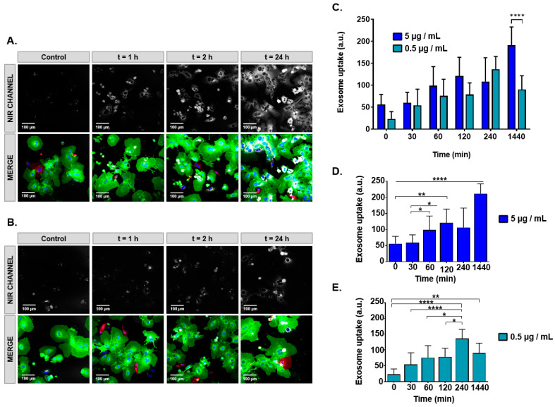
Figure 4.
Confocal microscope imaging for assessing the uptake of sulfo-cyanine 7.5-labeled milk exosomes (SCy-MiExo) by hepatocytes. (A,B) Near infrared (NIR, top) and fluorescence images (bottom) taken over time as hepatocytes internalized: (A) 5 µg/mL and (B); 0.5 µg/mL of SCy-MiExo; (C) Quantification of the exosome uptake at different SCy-MiExo concentrations, analyzed in regions of interest. Values were statistically significant between both concentration at 24 h; **** (p ≤ 0.0001). Data are expressed as the mean ± standard deviation. (D) Statistical analysis for the dose of 5 µg/mL: all values were significant compare to 24 h; * (p ≤ 0.05), ** (p ≤ 0.01), *** (p ≤ 0.001), **** (p ≤ 0.0001). (E) Statistical analysis for the dose of 0.5 µg/mL; * (p ≤ 0.05), ** (p ≤ 0.01), *** (p ≤ 0.001), **** (p ≤ 0.0001).

