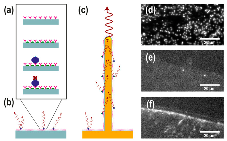
Figure 2.
(a) Schematic representation of the single chain fragment variable (scFv) assay used in this study. ScFv antibodies (pink) are physisorbed on a surface. A blocking agent (green) is subsequently used to minimize unspecific binding of biotinylated serum proteins (blue), which are targeted by fluorescently labelled streptavidin (dark red). (b,c) Schematic representation of the assay on a flat substrate (b) and a light-guiding nanowire substrate (c): the emission of fluorophores on the nanowire surface excites the supported waveguide modes to be reemitted at the tip, leading to an enhanced signal intensity at the tip of the nanowires. (d–f) Top-view epifluorescence images (displayed in the same pixel window) of (d) a GaP nanowire substrate, (e) a silicon substrate and (f) a MaxiSorp black polymer substrate, spotted with antibodies and exposed biotinylated serum concentration diluted to 0.4% (the highest concentration used in this study). Individual nanowires are visible in (d) as bright spots. The edge of the scFv spot is visible in (e,f), showing the difference in the signal between specific an unspecific binding to the antibodies.

