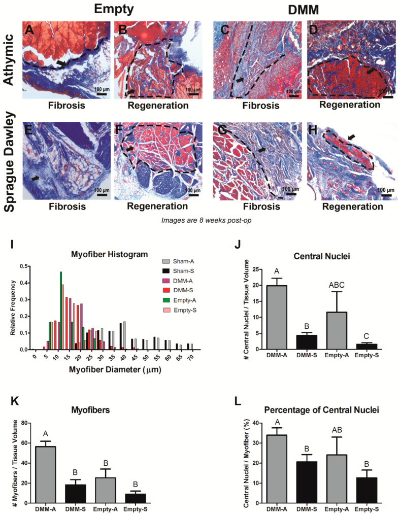
Figure 3.
Histological staining and morphometric analysis for VML injuries in Sprague Dawley and RNU rats. Representative images demonstrate a fibrotic response following injury or DMM treatment (A,C,E,G), or regenerative response following injury or DMM treatment (B,D,F,H). Histomorphometry showed similar relative frequencies for myofibers in Sprague Dawley and RNU rats (I), increased central nuclei in DMM-treated RNU rats (J), increased myofibers in DMM-treated RNU rats (K), and increased percentage of central nuclei in RNU rats (L). Letters not shared indicate a significant difference (p < 0.05, Tukey). Data shown are means ± SEM of 8 DMM and sham animals and ± SEM of 3 for empty defect animals.

